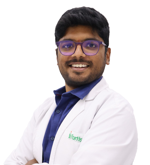
Diagnostic nuclear medicine uses radiopharmaceuticals and specialised cameras to examine how organs and tissues function. Dr. Chandra Teja Reddy Singareddy specialises in nuclear medicine imaging and molecular investigations. These techniques may help assess selected cancers, thyroid disorders and other medical conditions where functional imaging provides clinically useful information.
Positron Emission Tomography combined with Computed Tomography provides functional and anatomical information in a single imaging examination. Dr. Chandra Teja Reddy has expertise in PET-CT imaging and related research. The scan may be used for selected cancer investigations and other conditions depending on the tracer, clinical indication and diagnostic requirements.
FDG PET-CT uses a radioactive glucose analogue to identify tissues with increased metabolic activity. Dr. Chandra Teja Reddy has published research involving FDG PET-CT in various cancers. Imaging may assist with staging, treatment response evaluation and detection of selected abnormalities, although interpretation depends on clinical history and other investigations.
Gamma camera imaging detects radiation emitted by specific radiopharmaceuticals to produce information about organ function or disease activity. Dr. Chandra Teja Reddy has documented expertise in diagnostic gamma camera studies. Such examinations may support assessment of thyroid function and other conditions requiring specialised nuclear medicine investigations.
Radioactive iodine therapy uses iodine-131 to treat selected thyroid conditions. Dr. Chandra Teja Reddy has specialist training in therapeutic nuclear medicine and documented expertise in radioiodine treatment. Before treatment, patients undergo assessment of the thyroid diagnosis, relevant medical history and safety considerations, including pregnancy-related precautions and radiation protection requirements.
Radioiodine treatment may be recommended for selected patients with differentiated thyroid cancer, often following thyroid surgery. Dr. Chandra Teja Reddy has specific clinical and research expertise in radioactive iodine therapy for thyroid malignancies. Treatment decisions depend on cancer risk, previous treatment, radioiodine sensitivity and specialist recommendations.
Radioactive iodine can be used to treat certain benign thyroid disorders, particularly selected forms of hyperthyroidism. Dr. Chandra Teja Reddy is listed as having experience with radioiodine therapy for benign thyroid conditions. Patient suitability depends on the diagnosis, thyroid function, previous treatment and relevant medical contraindications.
Iodine-131 whole-body scanning is used in selected patients with differentiated thyroid cancer to identify iodine-avid thyroid tissue or disease. Dr. Chandra Teja Reddy has published research involving diagnostic and post-therapy iodine scans. Results may help specialists assess disease distribution and determine appropriate follow-up management.
Post-therapy radioiodine imaging is performed after selected radioactive iodine treatments to evaluate where the administered iodine has accumulated. Dr. Chandra Teja Reddy has investigated the additional value of post-treatment iodine scans in differentiated thyroid cancer. Findings may assist with subsequent disease assessment and treatment planning.
Nuclear medicine imaging can contribute to cancer staging by identifying selected sites of metabolically active or radiotracer-avid disease. Dr. Chandra Teja Reddy has clinical and academic experience in oncology-related PET-CT imaging. The choice of examination depends on cancer type, disease characteristics and multidisciplinary oncology recommendations.
PET-CT and other molecular imaging examinations may help evaluate treatment response in selected cancers. Dr. Chandra Teja Reddy has undertaken research on PET-CT imaging in breast cancer immunotherapy. Interpretation considers tracer uptake, prior imaging, treatment history and the possibility of inflammatory or treatment-related findings.
Neuroendocrine tumour imaging uses selected molecular tracers to identify tumours expressing relevant biological targets. Dr. Chandra Teja Reddy has contributed to research involving neuroendocrine tumour imaging, including Gallium-68 DOTANOC PET-CT. Such investigations may help assess disease distribution and support multidisciplinary decisions about further treatment.
Gallium-68 DOTANOC PET-CT is a molecular imaging technique used for evaluating selected somatostatin receptor-expressing neuroendocrine tumours. Dr. Chandra Teja Reddy has published research involving this tracer. The examination may assist with tumour localization, staging and assessment for certain targeted radionuclide treatments when clinically appropriate.
Parathyroid adenoma localization helps identify overactive parathyroid tissue in selected patients with primary hyperparathyroidism. Dr. Chandra Teja Reddy has contributed to research involving fluorocholine PET-CT for locating unusual parathyroid adenomas. Imaging findings may help surgeons plan treatment when conventional investigations are inconclusive.
Fluorocholine PET-CT uses a specialised radioactive tracer to examine selected metabolic processes. Dr. Chandra Teja Reddy has published research on its use in detecting parathyroid adenomas at unusual anatomical sites. Clinical use depends on the indication, tracer availability and suitability of the examination.
Yttrium-90 microsphere therapy is a targeted internal radiation treatment used for selected liver tumours. HexaHealth lists it among Dr. Chandra Teja Reddy's therapeutic nuclear medicine interests. Treatment usually requires coordinated assessment involving interventional radiology, nuclear medicine and oncology specialists, and its current availability must be confirmed with the hospital.